


Your First Pregnancy Scan
Finding out that you are expecting a baby can cause a whole mixture of emotions that range from excitement to worries and concerns as you embark on this new stage of your life. In England, you will be called for a pregnancy scan with the NHS at 12 weeks. We understand that you may be considering having a scan earlier to seek reassurance during those very early weeks.
We sat down with our women’s health sonographer Edel, who details what you could expect from an ultrasound scan during those early weeks from 4-7 weeks, to help you have better understanding if you are considering having an early pregnancy scan performed privately.
At our conveniently located private London clinics, our leading consultants and expert women’s health sonographers use state-of-the-art-imaging to carefully evaluate and look after you from the very first weeks up until 36 weeks.
| Gestational Age | What do we see on an ultrasound? |
| 4 weeks
|
In essence not much at all and this is normal.
We may see possible thickening of the lining of the womb (endometrium) in preparation for the implantation of pregnancy. |
| 5 weeks
|
A very early gestational sac within the endometrial cavity which is the first structure we see in pregnancy.
|
| 5.5 to 6 weeks
|
Within the gestational sac a yolk sac forms. The second structure that we see in pregnancy. The yolk sac provides nutrition between the mother and the developing embryo.
|
| 5.5 to 6.5 weeks
|
A fetal pole develops next to the yolk sac; It is sometimes possible to see a fetal heartbeat at this early stage. |
| 6.5 to 7 weeks
|
If you have regular menstrual cycles and are sure of your dates, by this stage we should see a clear gestation sac, yolk sac and fetal pole within the uterus. A fetal heartbeat can also be visualised at this stage. |
6 weeks ultrasound
The pregnancy is small at this early stage. The most we would expect to see is a gestation sac, yolk sac and fetal pole +/- a heartbeat. The fetal pole will look like a little pea on the screen (but hey, that is your special little pea!).
If we cannot see a heartbeat at this scan then this is either due to the pregnancy being too early (this is the most common reason) or that the pregnancy is found to be not viable.
What is the likely size of the fetus?
The fetus is anywhere between 2-7mm.
Any thoughts/advice for patients regarding a scan at 6 weeks /anything they should know?
If the lady has irregular periods or unsure of dates, we may not see anything on the scan at this point.
7 weeks ultrasound
By this stage we should see a clear gestation sac, yolk sac and fetal pole within the uterus. A fetal heartbeat can also be visualised at this stage. If we cannot see a heartbeat at this scan then this is either due to the pregnancy being too early (this is the commonest reason) or that the pregnancy is found to be not viable.
What is the likely size of the fetus?
Anywhere between 7-10mm.
Any thoughts/advice for patients regarding a scan at 7 weeks / anything they should know?
If you have irregular periods or are unsure of dates, we may not see all the structures at this point. This may be that you are less weeks pregnant than you calculated.
8 weeks ultrasound
By this stage, we should see a clear gestation sac, yolk sac and fetal pole within the uterus. A fetal heartbeat can also be visualised at this stage.
What is the likely size of the fetus?
Anywhere between 10-16mm. If we look closely, we may even start to see limb buds forming and slight movements.
Any thoughts/advice for patients regarding a scan at 8 weeks /anything they should know?
If you have irregular periods or are unsure of dates, we may not see all the structures at this point. This may be that you are less weeks pregnant than you calculated.
6-8 weeks ultrasound scans:
What will you be able to see /evaluate from a 6-8 weeks ultrasound scan?
From 6-8 weeks, we will be able to rule out ectopic pregnancy and check the pregnancy sac is located within the uterus. We will also be able to confirm the viability of pregnancy and check if there is a heartbeat present. We will also be able to calculate the gestation of pregnancy (number of weeks), and determine whether it is a singleton or multiple pregnancy. And look at any cause for bleeding/spotting or any unusual pain, in addition to an assessment of ovaries and pelvic area.
It’s very common for women to have a corpus luteum ovarian cyst during the first 3 months of pregnancy, which forms on the ovary where the egg is released, the sonographer will note side of the cyst during this scan.
What will happen during the appointment/scan?
To obtain clearer images of the early pregnancy (6-8 weeks), we offer an internal scan (vaginal) with your consent. This will not be harmful in any way to the baby. A trans-abdominal scan (over your tummy) will not be very beneficial at this early stage. As the pregnancy grows past 8-9 weeks, we can usually see over the tummy as long as you have a full bladder.
In our ultrasound room, there is a screen you can watch as the sonographer performs the scan and explains the findings to you.
What will happen after the ultrasound scan?
After the scan you will be provided with a full medical report and a photo print of your pregnancy.
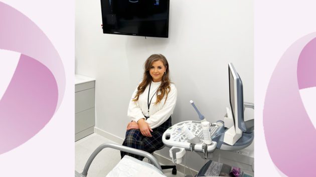
About Edel, expert women’s health sonographer at our city location
Edel is an experienced women’s health sonographer who joins our team from Southend University Hospital (NHS trust) where she is Deputy Ultrasound Manager and performs a large number of early pregnancy and emergency gynae scans. Edel is passionate about ensuring patients feel comfortable and at ease during appointments while treating them with compassion and kindness.
Edel confirms, “As a specialist sonographer, I have performed thousands of ultrasound scans for pregnancy. A lot of the time ultrasound scan findings are normal. However, if unexpected findings occur during your scan, such as miscarriage, then I will carefully communicate these and direct you to the necessary support you need.”
Make an appointment:
We are pleased to provide a variety of sonographer-led services at our modern and newly opened clinic in the historic Austin Friars Square in the City of London. The scan-only services include early pregnancy scanning (pre 13 weeks and fetal wellbeing scans) in addition to pelvic ultrasound scans. Prices start from £150, learn more here. To view availability and book, visit our online booking portal.